

Ultrasonographic Findings In Patients Diagnosed With Acute Appendicitis
Dr. Juan Pablo Ramírez


PURPOSE OR OBJETIVE:
Acute appendicitis is a frequent predominant pathology in pediatric ages. This pathology constitutes the main cause of acute surgical abdomen in emergencies. (1)
The early diagnosis of acute appendicitis is essential, it is generally through physical evaluation and clinical examination, however, the use of paraclinical studies, whether laboratory and imaging, help the surgeon to have a better pre-surgical vision, being more complete for each case and to have data that may be relevant to determine the approach or other relevant aspects to decide behaviors before, during and after the surgical act. Ultrasound is useful in all cases where acute appendicitis is suspected, especially when there is a diagnostic doubt based on the clinic or laboratory. (2)
Ultrasound can provide important information, especially in cases where there are doubts regarding the diagnosis, ultrasound can be a very useful tool for the surgeon.
It is important to highlight that the characteristics of the ultrasonographic images that we can find when evaluating a patient with acute appendicitis must be adequately known, in order to have greater precision when establishing a diagnosis. In this way, negative appendectomies can be reduced, reducing the morbidity and mortality inherent to this pathology and the complications related to the performance of unnecessary surgical acts. (3)
That is why the following questions are highlighted: ¿What are the ultrasonographic findings in patients with a diagnosis of acute appendicitis? And ¿What is the frequency of ultrasonographic findings found in patients with a clinical diagnosis of acute appendicitis?
In this way, the following learning objectives are proposed:
Overall objective:
Describe the ultrasonographic findings in patients diagnosed with acute appendicitis.
Specific objectives:
Determine which are the most frequent ultrasonographic findings in patients diagnosed with acute appendicitis.
Characterize the ultrasonographic findings in patients diagnosed with acute appendicitis.
Establish the frequency of each of the ultrasonographic findings evidenced in patients diagnosed with acute appendicitis.
MATERIALS AND METHODS:
Type of study:
Descriptive, prospective cross-sectional study.
Shows:
The sample was made up of all patients with clinical suspicion of acute appendicitis, who underwent abdominal ultrasound, in the Advanced Ultrasound and Vascular Doppler Service of “El Paso” Teaching Medical Center (AUVDSEPTMC), since August from 2019 to October 2020, which were confirmed by the surgical act and pathological anatomy. The sample is intentional, not probabilistic, limited to the number of cases presented in the study period, corresponding to 12 patients.
Inclusion criteria:
Patients with clinical suspicion of acute appendicitis, who underwent abdominal ultrasound and with surgical and pathological confirmation of the diagnosis.
Patients with approval of their participation in the study by means of informed consent.
Exclusion criteria:
Patients whose diagnosis of acute appendicitis was not confirmed in the surgical act or by pathological anatomy.
Patients who did not wish to participate in said study by refusing to sign the informed consent.
Patients evaluated outside the study period.
METHOD
Twelve patients who met the inclusion criteria were admitted to the study, who presented a clinical diagnosis of acute appendicitis, to whom an abdominal ultrasound was requested at the Advanced Ultrasound and Vascular Doppler Service of “El Paso” Teaching Medical Center (AUVDSEPTMC), from August 2019 to October 2020. Abdominal ultrasound was performed using a “Medison” Brand Ultrasound, Model “Acuvixx”, with Convex and linear multifrequential transducers. When performing the abdominal ultrasound, the due report was reported, each of the findings found were characterized, which were later compiled in "The data collection sheet of patients with a clinical diagnosis of acute appendicitis evaluated by ultrasound", later these diagnoses were confirmed by the surgical and pathological findings. Likewise, later the patient data were compiled and characterized using the Microsoft Excel program.
RESULTS:
Twelve patients with a diagnosis of acute appendicitis, later confirmed by surgical and pathological findings, were evaluated by abdominal ultrasound, eight (66.7%) were men and four women (33.3%), with an age range between 5 and 50 years, with an mean of 25.9 years.
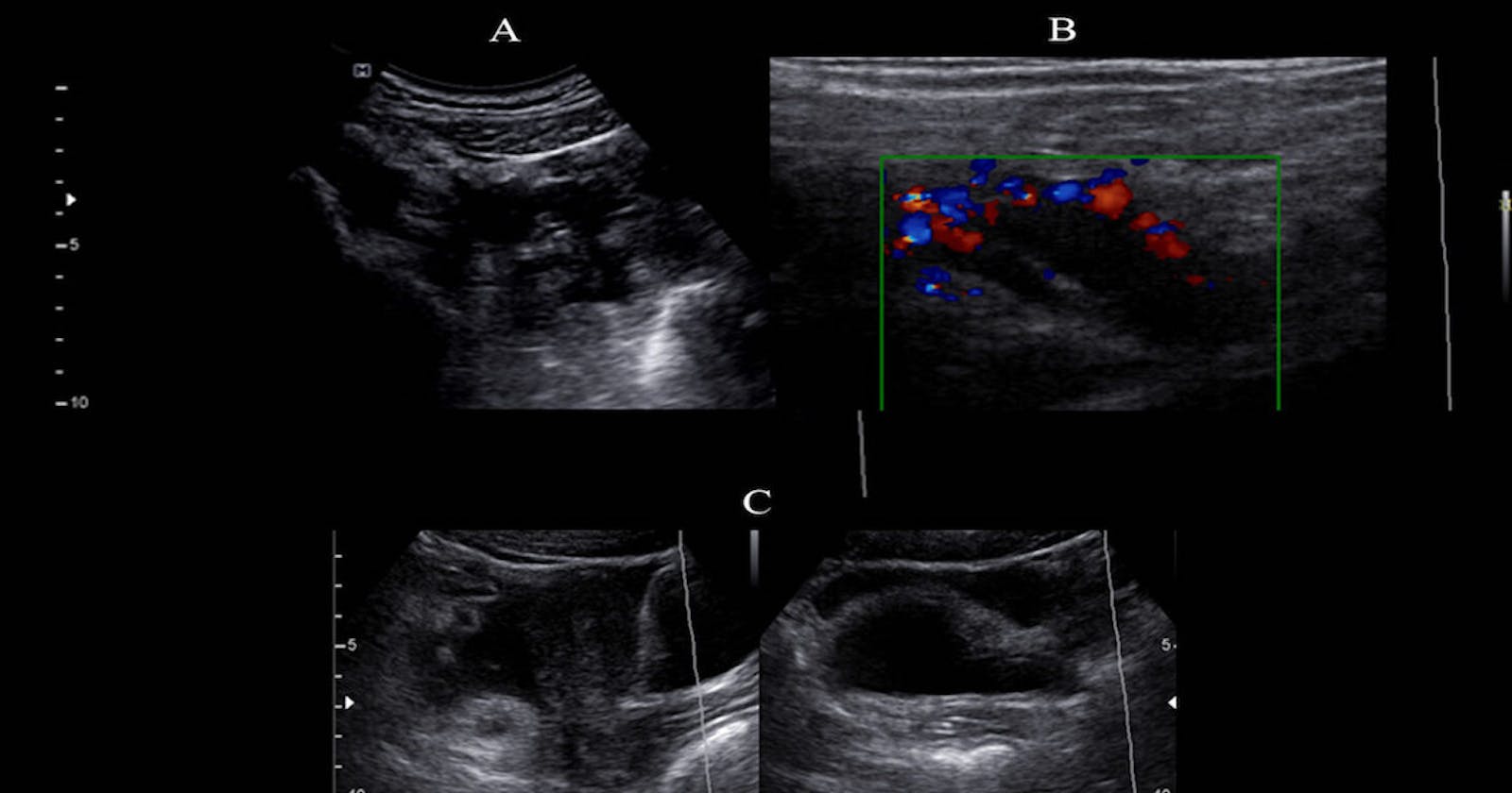
The findings found in 12 (100%) of the patients diagnosed with acute appendicitis were the absence of compressibility, the glove finger sign and the target sign. These being the most frequently found findings. (See Figure 1)
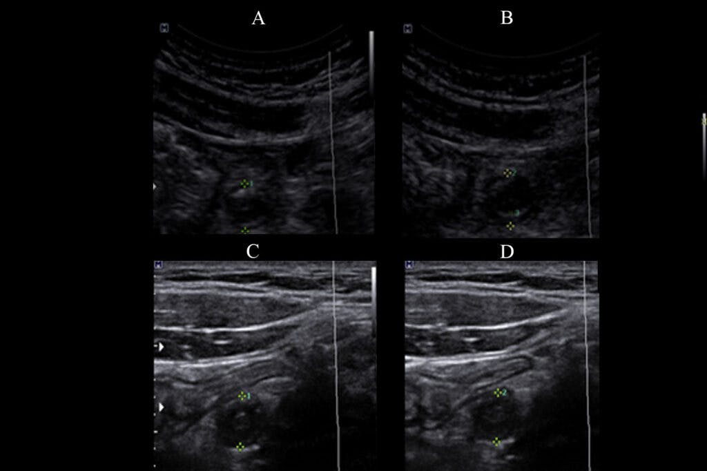
Fig 1: "A" and "B" show the ultrasound visualization of the appendix in a cross section, in a patient with a diagnosis of acute appendicitis, without compression and when compression with the transducer, note the absence of compressibility of the appendix, as well as in the images labeled "C" and "D", corresponding to another patient.
The glove finger sign describes the visualization of a tubular structure emerging from the cecum. While the sign of the target is given by the circular appearance of the appendix in the orthogonal plane. The absence of compressibility is also described as a direct ultrasound sign suggestive of acute appendicitis.(4)(See Figure 2 and Figure 3).
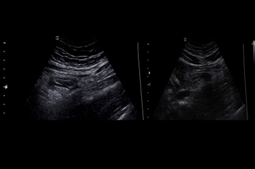
Fig 2: Two images are presented corresponding to the ultrasound visualization of the appendix in longitudinal axis, of two patients with a diagnosis of acute appendicitis. A tubular image with a blind background can be seen, without the presence of peristaltic movements that determine the sign of the “glove finger”.
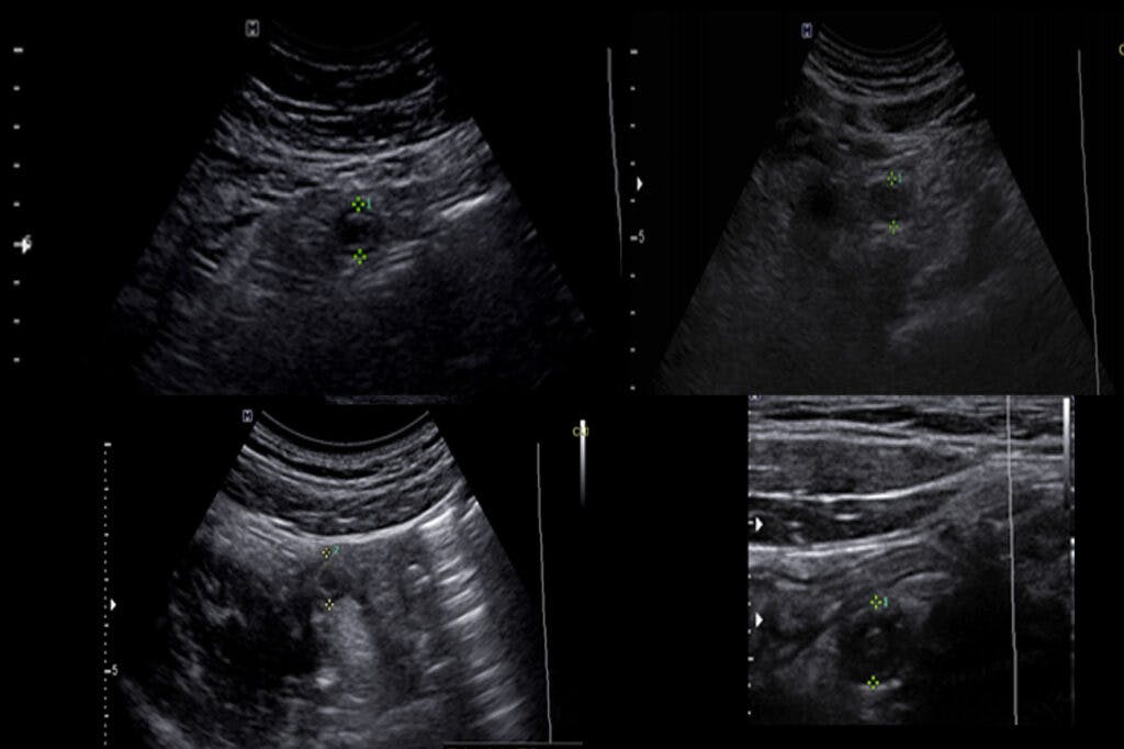
Fig 3: Four images corresponding to the ultrasound visualization of the appendix in a transverse plane are displayed, where the circular arrangement of its layers is appreciated, determining the “target sign”, as well as the alteration of the adjacent fat.
The thickening of the appendix wall (greater than 3 mm) and the increase in the diameter of the appendix (greater than 7 mm) was evidenced in 83.3% of the sample. Some authors refer to a diameter of the appendix in orthogonal section greater than 6 mm, however taking this value also influences the 7 mm considered in the study. (4,5) (See Figure 4).
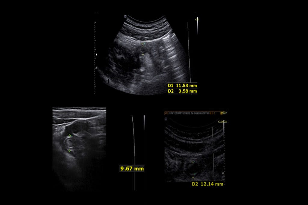
Fig 4: A cross section of the appendix of three patients with acute appendicitis is visualized sonographically. Note the appendicular diameter greater than 7 mm and the wall thickness greater than 3 mm.
The alteration of the adjacent mesenteric fat of the appendix was visualized with a frequency of 66.7%. The increased echogenicity of locoregional fat is a suggestive finding of an acute appendicular process. (5)
Fluid in the right parietocolic slide was seen in 33.3% of the evaluated patients. The presence of fluid is considered an indirect ultrasound finding for the diagnosis of acute appendicitis.(4,5)(See Figure 5_a).
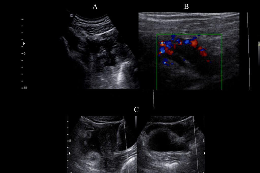
Fig 5: Three images of additional findings found in patients with acute appendicitis are displayed: A) Scant amount of fluid in the right parietocolic gutter. B) Increased flow uptake to the color Doppler signal from the appendix. C) Presence of liquid in the cul-de-sac.
Increased flow uptake to the color Doppler signal of the appendix was found in 25% of cases, this ultrasound finding is also considered an indirect finding for the diagnosis of acute appendicitis.(4) (See Figure 5_b).
Liquid was observed in the cul-de-sac and the presence of gas in the appendix wall in 8.3% of the cases. (See Figure 5_c).
CONCLUSION:
The ultrasonographic findings found in patients with a clinical diagnosis of acute appendicitis, confirmed by surgical procedure and pathological anatomy, were: the absence of compressibility of the appendicular lumen, the glove finger sign, the target sign, the increase in thickness of the appendicular wall greater than 3 mm, the increase in the diameter of the appendix greater than 7 mm, alteration of the mesenteric fat adjacent to the appendix, increased uptake of flow to the color Doppler signal of the appendix, the presence of gas in the wall appendicular and the presence of fluid in the right parietocolic gutter and / or sack bottom.
The most frequent ultrasound findings in patients diagnosed with acute appendicitis are: the absence of compressibility of the appendix, the glove finger sign and the target sign, being found in 100% of patients diagnosed with acute appendicitis, followed by a increase in the thickness of the appendicular wall (greater than 3 mm) and an increase in the appendicular diameter (greater than 7 mm), seen in 83.3% of patients. (See Figure 6).
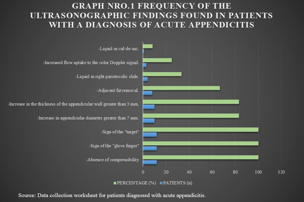
Fig 6: Graph Nro.1 Frequency of the ultrasonographic findings found in patients with a diagnosis of acute appendicitis. Note that the most frequently found findings in all patients was the absence of compressibility, the glove finger sign and the target worthy. Followed by an increase in the appendicular diameter and an increase in the thickness of the appendicular wall.
The ultrasound findings that occurred less frequently were the presence of fluid in the cul-de-sac and the presence of gas in the appendicular wall.
The use of ultrasound to complement the diagnosis of acute appendicitis initially based on the clinical manifestations and signs provides important information for the surgeon, being of greater relevance when there are doubts regarding the diagnosis, in these cases ultrasound can provide data that help reduce the possibilities of negative appendectomies, the identification of complications or the determination of the presence of differential diagnoses, guiding the surgeon to take the most appropriate behaviors for the management of each case.
REFERENCES:
Velásquez Hawkins C, et al. Value of ultrasound in the diagnosis and management of acute appendicitis. Rev Gastroenterol Peru; 27: 259-263. scielo.org.pe/pdf/rgp/v27n3/a06v27n3.pdf
Margain PMÁ, Vera RF, Dimas UN. Ultrasound for the diagnosis of appendicitis at Hospital Angeles Metropolitano. Acta Med. 2014; 12 (2): 65-70.
Arevalo Espejo O, et al. Acute appendicitis: Imaging Findings and current approach to diagnostic imaging. Rev Colomb Radiol. 2014; 25 (1): 3877-88. webcir.org/revistavirtual/articulos/noviemb..
Martinez Moya M, et al. Acute appendicitis. Multidisciplinary Study. SERAM 2008. Poster: seram2008.seram.es/modules.php?name=posters..\=
Peris Perez M.L, et al. Evaluation of ultrasound as a diagnostic method in emergencies of acute appendicitis: Our experience. dx.doi.org/10.1594/seram2014/S-0458